
Abstract
Objectives
Targeting drug-metabolizing enzymes in tumor cells may improve chemoprevention and chemotherapy by providing new targets for anticancer agents or combination therapies. This study investigated whether inhibition of CYP1A1, CYP3A4, and GSTP1 by α-naphthoflavone, ketoconazole, and quercetin modulates docetaxel activity in PC3 prostate cancer cells and PNT1A prostate epithelial cells.
Materials and Methods
PC3 and PNT1A cells were exposed to docetaxel either alone or in combination with inhibitors of CYP1A1, CYP3A4, and GSTP1. Sub-lethal monotherapy doses were calibrated to produce approximately 70% cell viability at 24 hours and 60% at 48 hours. Cell viability was assessed by WST-1 assay, while apoptosis and necrosis were evaluated using Annexin V staining. Enzyme activities were measured by ELSA. Cellular oxidative activity was assessed by DCFDA fluorescence analysis.
Results
In PC3 cells, calibrated monotherapy doses were: 4 µM docetaxel, 1000 nM α-naphthoflavone, 56.5 µM ketoconazole, and 300 µM quercetin. Combination treatments reduced cell viability by an additional 12–25% at 24 hours and by up to 45% at 48 hours, compared with docetaxel alone. Docetaxel treatment induced time-dependent changes in enzyme activities, whereas combination treatments markedly reduced DCFDA fluorescence signals.
Conclusion
Co-administration of docetaxel with CYP1A1, CYP3A4, and GSTP1-related pharmacological modulators may enhance cytotoxicity in prostate cancer cells. Increased cytotoxicity was accompanied by reduced DCFDA fluorescence, indicating that enhanced treatment responses were not necessarily associated with increased oxidative fluorescence signals.
Keywords: Prostate cancer, docetaxel, biotransformation enzymes, enzyme inhibitors
INTRODUCTION
Prostate cancer (PCa) is a major health concern among men and contributes substantially to cancer-related mortality worldwide. Based on their response to androgenic stimulation, prostate tumors are generally categorized into androgen-dependent forms, which require testosterone for growth, and castration-resistant or androgen-independent forms, which continue to progress even when testosterone levels are suppressed to castrate concentrations. Although androgen suppression therapy (AST) is initially effective, disease progression frequently leads to metastatic castration-resistant prostate cancer (mCRPC), for which treatment options remain limited. FDA-approved treatments for mCRPC include docetaxel, abiraterone, enzalutamide, cabazitaxel, and sipuleucel-T;,,,, however, their limited survival benefits highlight the need for novel therapeutic strategies, including combinations of chemotherapy, immunotherapy, radiotherapy, and AST. Docetaxel is widely used to treat several solid tumors, including PCa., Hepatic metabolism of DTX occurs predominantly via cytochrome P450 (CYP) enzymes, particularly CYP3A4 and CYP3A5, and CYP3A4 is considered a major determinant of interpatient differences in DTX clearance.,, CYP3A4 metabolizes docetaxel into inactive hydroxylated metabolites, reducing its therapeutic efficacy., CYP1A1 also contributes to drug metabolism and drug–drug interactions, including docetaxel metabolism. In Phase II metabolism of docetaxel, GSTP1 mediates the glutathione conjugation of docetaxel metabolites., According to Murray et al.,CYP3A protein is not detected in normal prostate tissue but expressed in 61% of prostate tumor specimens. They also observed the expression of CYP1A (63%), CYP2C (25%), epoxide hydrolase, and various GST isoforms in non-neoplastic prostatic epithelium. Furthermore, GSTP1 overexpression has been linked to reduced antitumor activity. Compared with androgen-dependent LNCaP cells, PC3 cells exhibit a significantly higher expression level, suggesting that it may contribute to the proliferative activity of androgen-independent PCa cells.
Modulation of enzymes involved in drug metabolism in tumor cells may improve chemoprevention and chemotherapy by offering new targets for anticancer agents or for combination therapies., Among the biotransformation-related enzymes of interest, CYP3A4 plays an established role in docetaxel metabolism, whereas CYP1A1 and GSTP1 may influence drug responses through xenobiotic metabolism and detoxification. Accordingly, ketoconazole and α-naphthoflavone were selected to modulate the Phase I enzymes CYP3A4 and CYP1A1, respectively, and quercetin was selected for its reported inhibitory activity toward GSTP1, a Phase II enzyme.,, This study investigated the effects of sub-lethal doses of these compounds, combined with docetaxel, on PC3 PCa cells and PNT1A non-malignant prostate epithelial cells by assessing cell viability, apoptosis, enzyme activities, and cellular oxidative activity. The findings may provide insight into biotransformation-related pathways affecting docetaxel responsiveness and inform future therapeutic strategies.
MATERIALS AND METHODS
WST-1 cell proliferation assay
Cell viability was assessed using the WST-1 assay. Cells were seeded in 96-well plates (10,000 cells/well) and treated in triplicate with docetaxel, quercetin, α-naphthoflavone, or ketoconazole in RPMI medium; DMSO was used as the vehicle control. After 24 or 48 hours, 50 µL of WST-1 reagent was added to each well, and absorbance was measured using a SpectraMax Paradigm Multi-Mode Plate Reader, as described by Nayman et al.
Protein isolation
Following 24-hour docetaxel treatment, PC3 and PNT1A cells were scraped into a manually prepared lysis buffer and whole-cell lysates were sonicated as previously described. Total protein concentrations were determined using the Pierce Coomassie Protein Assay Kit (Thermo Scientific, USA) according to the manufacturer’s instructions.
Enzyme activities
PC3 and PNT1A lysates collected after 24 or 48 hours of docetaxel exposure were used to determine the enzymatic activities of total GST, GSTP1, CYP1A1, and CYP3A4. Total GST activity was determined with CDNB/GSH, whereas GSTP1 activity was measured using ethacrynic acid/GSH, both following the protocol reported by Habig et al., CYP1A1 activity was assessed using the EROD assay, as optimized by Sen and Arinc based on the original method described by Burke and Mayer. CYP3A4 activity was measured using the erythromycin N-demethylation assay, in which formaldehyde formation was quantified according to Nash’s method as modified by Cochin and Axelrod. Enzyme activities were normalized to protein content.
Annexin-V FLUOS staining assay
PC3 cells were treated with CCG inhibitors alone or in combination with docetaxel for 24 hours. Apoptosis was assessed using the Annexin V-FLUOS staining kit (Roche, 11988549001) according to the manufacturer’s instructions, as previously described by Nayman et al. The apoptotic cell population was determined by flow cytometric analysis using a BD FACSCalibur flow cytometer (Becton, Dickinson and Company, USA).
Analysis of ROS activity of quercetin, α-naphthoflavonoid, and ketoconazole in PCa PC3 Cells
To evaluate ROS activity, PC3 cells were seeded into 96-well plates at 2.5 × 10 cells/well and treated with sub-lethal doses of CCG inhibitors, docetaxel, or docetaxel-based combinations. Following 48 hours of exposure, cells were washed with 1 × reaction buffer and then incubated in the dark with DCFDA solution at 37 °C for 1 hour. After washing once more, ROS activity was determined using fluorescence microscopy.
Statistical analysis
GraphPad Prism 11 was used to perform the statistical analyses. Prior to hypothesis testing, normality and log-normality were checked, and the Shapiro–Wilk test was used to assess normality. Homogeneity of variance was tested with the Brown–Forsythe test. Welch’s t-test was selected for two-group analyses when variances were unequal, while Welch’s analysis of variance (ANOVA) was used for comparisons among multiple groups when variances were unequal. For experiments with two independent variables, two-way ANOVA was performed, followed by suitable multiple comparison tests when applicable. Results are reported as mean ± standard deviation, with statistical significance defined as p < 0.05.
RESULTS
Establishing sub-lethal concentrations for docetaxel and CCG inhibitors
To determine whether CCG inhibitors enhance docetaxel efficacy, sublethal concentrations were first established to permit assessment of the effect of the combination treatment. Concentrations causing approximately 30% reduction in viability for CCG inhibitors and a 50% reduction in viability for docetaxel at 48 hours were selected to avoid maximal cytotoxicity and to enable combination analysis (). The WST-1 assay was used to evaluate the effects of docetaxel and CCG inhibitors on PNT1A and PC3 cell viability. In PC3 cells, single-agent treatments with 4 µM docetaxel, 1000 nM α-naphthoflavone, 56.5 µM ketoconazole, or 300 µM quercetin each reduced viability to approximately 70% after 24 hours and to approximately 60% after 48 hours (–1D). Accordingly, combining docetaxel with CCG inhibitors at the selected concentrations further reduced PC3 cell viability compared with docetaxel alone. At 24 hours, co-treatments with α-naphthoflavone, ketoconazole, and quercetin produced additional reductions in viability of approximately 15%, 20%, and 17–18%, respectively; at 48 hours, the reductions were more pronounced, reaching approximately 15%, 30–33%, and 40–45%, respectively (). Importantly, treatment with CCG inhibitors at these concentrations did not significantly alter the viability of PNT1A control cells (), suggesting differential cellular sensitivity. Collectively, these results demonstrate that CCG inhibitors enhance the cytotoxic effect of docetaxel in a time-dependent manner, with ketoconazole and quercetin showing the most pronounced potentiation at 48 hours.
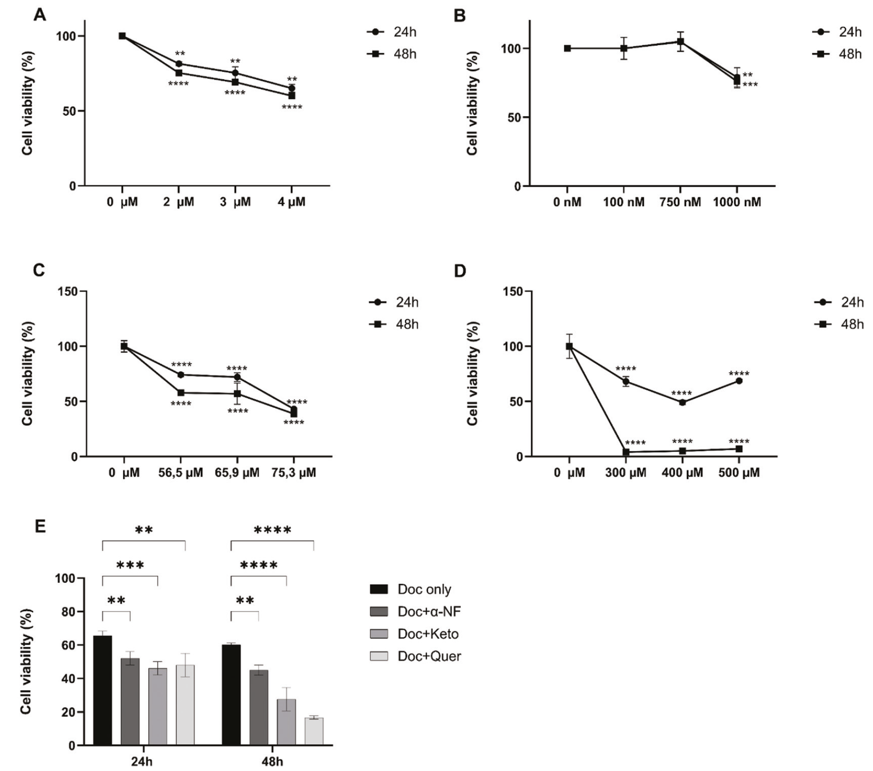
Effects of CCG inhibitors and combination regimen on cell viability. Effect of only Doc (A) or only CCG inhibitors on PC3 cell cell viability [CCG: α-NF (B), Keto (C) and Quer (D)] and combination regimen (E). Data are expressed as mean ± SD (n = 3). 100% viability is adjusted by taking the absorbance value obtained for vehicle-tratedtreated cells (control).
SD, standard deviation; α-NF, α-naphthoflavone; Keto: ketoconazole; Quer, quercetin; Doc, docetaxel; p ≤ 0.05; *p ≤ 0.01; ***p ≤ 0.001, ****p ≤ 0.0001.
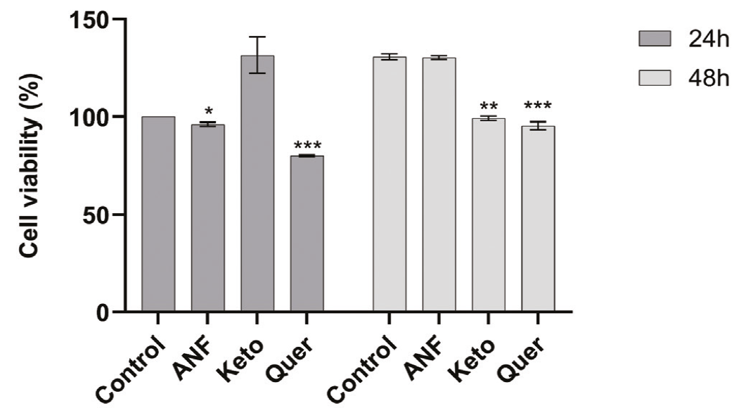
Effects of α-NF, Keto and Quer on PNT1A cell viability. Data are expressed as mean ± SD (n = 3). p ≤ 0.05; *p ≤ 0.01; ***p ≤ 0.001.
SD, standard deviation; α-NF, α-naphthoflavone; Keto, ketoconazole; Quer, quercetin.
Evaluating the effects of docetaxel on drug-metabolizing enzymes in PC3 cells
We next investigated whether docetaxel altered enzymatic activities in PC3 cells. Docetaxel treatment produced distinct, time-dependent alterations in drug-metabolizing enzyme activities (). In particular, CYP1A1 activity decreased by 2.6-fold at 24 hours, but then showed a marked 8.9-fold increase at 48 hours compared with the control group. CYP3A4 activity increased 1.3-fold at 24 hours and showed no significant change at 48 hours. Total GST activity remained unchanged at 24 hours but increased to 1.7-fold at 48 hours, while GSTP1 activity decreased to 0.8-fold at 24 hours and increased to 1.2-fold at 48 hours. Overall, these findings indicate an early suppression followed by compensatory upregulation of metabolic enzyme activities after docetaxel treatment.
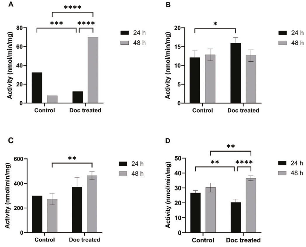
Effects of docetaxel treatment on CYP1A1 (A), CYP3A4 (B), total GST (C), and GSTP1 (D) enzymatic activities in PC3 cells. Data are expressed as mean ± SD (n = 3).
Doc, Docetaxel; p ≤ 0.05; *p ≤ 0.01; ***p ≤ 0.001; ****p ≤ 0.0001.
Assessing the effects of docetaxel and CCG inhibitors on apoptosis in PC3 cells
To assess whether combinations of docetaxel and CCG inhibitors enhance apoptosis in PC3 cells, Annexin V–PI staining was conducted after 24-hour treatments. Docetaxel markedly increased early apoptosis to 70%, whereas α-naphthoflavone, ketoconazole, and quercetin, when used alone, induced early apoptosis to 61%, approximately 45%, and 31%, respectively.
In combination, ketoconazole + docetaxel further increased early apoptosis to 83% (~38% increase compared with ketoconazole alone). In contrast, α-naphthoflavone + docetaxel showed comparable levels (~60–64%), indicating no additional effect. Quercetin plus docetaxel increased early apoptosis to 62% (~2-fold increase compared with quercetin alone).
Late apoptotic cell death was also elevated after docetaxel treatment, increasing from 0% in the control group to 18%. α-naphthoflavone alone increased late apoptosis to 27%, and late apoptosis increased further to 32% when α-naphthoflavone was combined with docetaxel. Quercetin treatment induced late apoptosis in 36% of cells, which increased to 40% in the combination group. By contrast, ketoconazole, either alone or in combination with docetaxel, induced lower levels of late apoptosis, ranging from approximately 12% to 16% (). Collectively, these findings show that CCG inhibitors differentially modulate docetaxel-induced apoptosis in PC3 cells. Ketoconazole mainly enhanced early apoptosis, while α-naphthoflavone modestly increased late apoptosis without affecting early apoptosis. Notably, quercetin markedly potentiated early apoptosis with only a modest effect on late apoptosis, suggesting preferential enhancement of apoptosis initiation rather than progression.
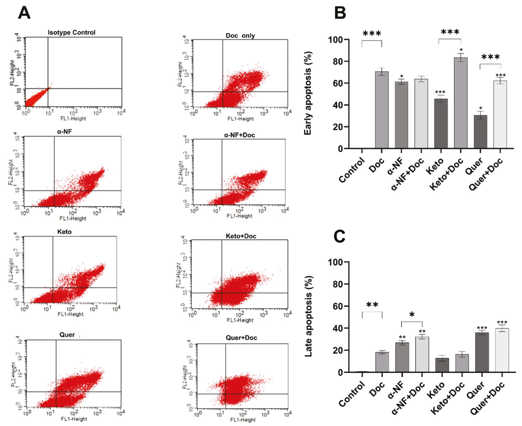
Effects of docetaxel, inhibitors, and combination treatments on cell death in PC3 cells. Representative flow cytometry analysisflow cytometric analysis of PC3 cell death is shown in panel (A) statistical analysis of flow cytometry data for early apoptosis (B) and late apoptosis (C), obtained after 24-hour treatment at the indicated concentrations, was performed using FITC–Annexin V staining. Data are expressed as mean ± SD (n = 3).
Doc, docetaxel; α-NF, α-naphthoflavone; Keto, ketoconazole; Quer: quercetin; p ≤ 0.05; *p ≤ 0.01; ***p ≤ 0.001. (The p-values for each inhibitor correspond to comparisons with docetaxel-only treatment).
Investigating the effects of docetaxel and CCG inhibitors on ROS-associated DCFDA fluorescence in PC3 cells
To determine whether CCG inhibitors modulate docetaxel-induced oxidative stress in PC3 cells, intracellular ROS levels were quantified by fluorescence intensity analysis. Docetaxel increased DCFDA fluorescence from 61% in untreated controls to 77%, whereas quercetin, α-naphthoflavone, and ketoconazole, when used alone, reduced fluorescence to 20%, 14%, and 21%, respectively. Combination treatments further decreased DCFDA fluorescence to 6,1%, 7,2%, and 19,7% for quercetin + docetaxel, α-naphthoflavone + docetaxel, and ketoconazole + docetaxel, respectively. Overall, all three CCG inhibitors reduced DCFDA fluorescence; the greatest reductions occurred in the quercetin + docetaxel and α-naphthoflavone + docetaxel groups ().
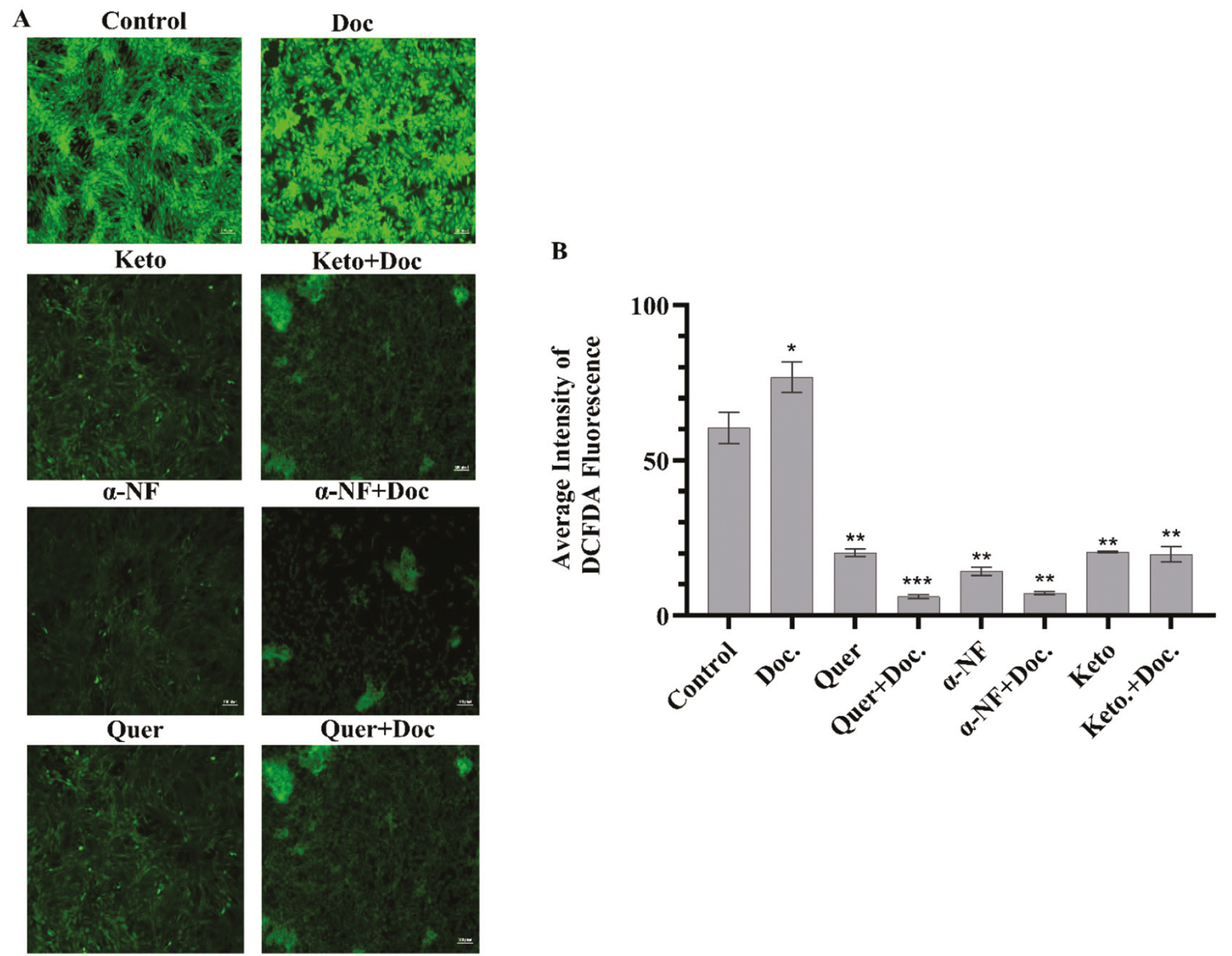
DCFDA fluorescence in PC3 cells following treatment with docetaxel and antioxidant compounds. (A) representative fluorescence microscopy images showing intracellular DCFDA fluorescence signals in PC3 cells treated with quercetin (300 µM), α-naphthoflavone (1 µM), ketoconazole (56.5 µM), and docetaxel, either alone or in combination, compared with the untreated control cells (CNT). Scale bar: 100 µm. (B) quantification of DCFDA fluorescence intensity in PC3 prostate cancer cells after 48-hour treatment with docetaxel (Doc), α-naphthoflavone (α-NF), ketoconazole (Keto), quercetin (Quer), and their corresponding combinations. Data are expressed as mean ± SD (n = 3). Statistical significance was assessed relative to the control group. p < 0.05; *p < 0.01; ***p < 0.001.
α-NF, α-naphthoflavone; Keto, ketoconazole; Quer: quercetin; Doc, docetaxel.
DISCUSSION
Docetaxel-based regimens are widely used as first-line chemotherapy for metastatic PCa; however, their efficacy is limited by chemoresistance and dose-dependent toxicity. Docetaxel resistance involves multiple cellular and molecular alterations, including emerging evidence of extracellular RNA-mediated modulation of drug responsiveness.,, Therefore, docetaxel-based combination strategies are being explored to improve therapeutic efficacy in PCa., Phase II trials showed that docetaxel plus estramustine improved response rates but increased toxicity, while subsequent Phase III trials evaluated whether this combination offered a meaningful clinical advantage in androgen-independent PCa. Based on this rationale, three pharmacological agents were selected to investigate the potential role of biotransformation-related pathways in the cellular response to docetaxel. α-naphthoflavone and ketoconazole were selected for their reported inhibitory effects on the Phase I enzymes CYP1A1 and CYP3A4, respectively, whereas quercetin was included for its inhibitory activity toward GSTP1, a Phase II detoxification enzyme.,, The present study aimed to explore how these compounds may affect the response of PCa cells to docetaxel by evaluating cell viability, apoptosis-associated changes, enzyme activities, and cellular oxidative activity.
Our results demonstrated treatment- and time-dependent changes in PC3 metabolic viability following combination treatments with docetaxel, with more pronounced effects observed with ketoconazole and quercetin than with α-naphthoflavone, particularly at later time points. These findings suggest that the tested inhibitors, particularly quercetin, may enhance docetaxel cytotoxicity through pharmacokinetic and pharmacodynamic mechanisms.,, Ketoconazole and α-naphthoflavone may influence docetaxel responsiveness through CYP inhibition, potentially affecting drug exposure,, whereas quercetin exhibits intrinsic antitumor properties—modulating survival signaling and promoting apoptosis—that may contribute to enhanced docetaxel sensitivity in PCa cells., As a result, quercetin–docetaxel co-treatment may produce enhanced cytotoxic effects, potentially improving therapeutic efficacy and influencing the development of drug tolerance. The stronger effect of quercetin at 48 hours may involve modulation of survival-signaling and drug-efflux mechanisms, potentially prolonging docetaxel-induced cytotoxicity.,
An important consideration was the cytotoxicity of the selected inhibitors in non-malignant prostate epithelial cells. At the tested concentrations, α-naphthoflavone, ketoconazole, and quercetin did not significantly affect PNT1A cell viability (); however, their combinations with docetaxel reduced PC3 cell viability more markedly than docetaxel alone (), suggesting differential cellular sensitivity. Although these compounds have been reported to inhibit CYP1A1, CYP3A4, and GSTP1, their broader pharmacological effects may also contribute to the enhanced docetaxel responsiveness observed in PC3 cells.
The temporal changes in the activities of CYP1A1, CYP3A4, total GST, and GSTP1 indicate alterations in biotransformation-related enzyme activities following docetaxel exposure. An approximately 2.6-fold decrease in CYP1A1 enzymatic activity at 24 hours, followed by an 8.9-fold increase at 48 hours, compared with the control group, indicates a pronounced time-dependent response to docetaxel exposure. This biphasic pattern may reflect an initial suppression of CYP1A1 activity followed by an adaptive cellular response during prolonged drug exposure. CYP1A1 expression is primarily regulated through the aryl hydrocarbon receptor (AhR) signaling pathway, which responds to endogenous ligands and xenobiotic compounds. Furthermore, oxidative stress, cellular stress-associated signaling pathways, and post-transcriptional regulatory mechanisms can influence CYP1A1 expression and enzymatic activity.,,, In this context, the marked increase in CYP1A1 activity at 48 hours may represent a compensatory response aimed at restoring cellular metabolic homeostasis following prolonged docetaxel exposure. Collectively, these findings suggest that CYP1A1 activity is dynamically regulated during docetaxel treatment, potentially reflecting time-dependent changes in the cellular response to chemotherapeutic stress. In contrast, CYP3A4 activity increased at 24 hours, potentially reflecting an early detoxification response, consistent with the reported role of PXR- and CAR-mediated CYP3A4 regulation in docetaxel metabolism and chemotherapeutic resistance. The increase in total GST activity, together with significant GSTP1 upregulation at 48 hours, may reflect activation of Nrf2-regulated antioxidant and Phase II detoxification pathways in response to chemotherapy-associated oxidative and electrophilic stress., Notably, GSTP1 contributes to taxane resistance by neutralizing drug intermediates and suppressing JNK-dependent apoptosis, while its late-phase induction may indicate a protective, drug-tolerant phenotype., Collectively, these time-dependent enzymatic alterations may reflect adaptive remodeling of xenobiotic metabolism and redox defense systems, potentially contributing to reduced docetaxel efficacy and chemoresistance in PCa.
To investigate the enhanced cytotoxicity of combination treatments, apoptosis was evaluated in PC3 cells using Annexin V assays. Docetaxel, alone or in combination with CCG inhibitors, increased the early and late apoptotic populations, consistent with its microtubule-stabilizing activity, induction of G2/M arrest, and activation of intrinsic apoptotic signaling. Docetaxel has also been reported to enhance apoptosis in PCa cells by sensitizing them to TRAIL through epigenetic upregulation of DR4 in vitro. Importantly, a further increase in apoptotic fractions observed with docetaxel–CCG inhibitor combinations suggests that these agents potentiate programmed cell death rather than inducing non-specific cytotoxic damage. The distribution of early and late apoptotic populations, however, varied among the combination treatments and may have reflected differences in the pharmacological properties of the selected inhibitors. α-naphthoflavone modulates CYP1A1 activity and AhR-related signaling, which may influence cellular responses to chemotherapeutic stress. In contrast, ketoconazole inhibits CYP3A-mediated metabolism, whereas quercetin has been reported to modulate survival-associated pathways, potentially influencing cellular susceptibility to apoptosis. These differences in pharmacological activity may contribute to the distinct apoptotic profiles observed following combination treatment. Specifically, the combination of α-naphthoflavone and docetaxel was associated with a greater late-apoptotic fraction, whereas combinations of ketoconazole and quercetin predominantly affected early-apoptotic populations. Nevertheless, Annexin V/PI staining provides a snapshot of apoptosis-associated cellular changes, and the observed distributions cannot independently establish differences in apoptotic progression or the involvement of specific molecular pathways., Thus, the differential apoptotic responses may reflect the combined influence of biotransformation-related enzyme modulation and other pharmacological activities of the selected compounds. Previous studies suggest that quercetin enhances docetaxel sensitivity through androgen receptor and PI3K/Akt signaling and caspase-dependent apoptosis,, whereas ketoconazole increases docetaxel exposure by inhibiting CYP3A-mediated metabolism., Collectively, these findings suggest that CCG inhibitors may enhance docetaxel-induced apoptosis, warranting further investigation of their potential to improve docetaxel responsiveness in PCa. However, apoptotic responses did not always correlate with WST-1 results, likely reflecting differences in assay endpoints: WST-1 measures cellular metabolic activity, whereas Annexin V/PI staining detects phosphatidylserine externalization and changes in membrane integrity., Although docetaxel combinations reduced metabolic viability in PC3 cells, these reductions were not consistently reflected in early and late apoptotic populations, indicating that changes in metabolic viability do not necessarily correspond to proportional changes in apoptosis. Moreover, WST-1 was assessed at 24 and 48 hours, whereas Annexin V/PI analysis was limited to 24 hours, restricting direct comparisons due to differences in assay endpoints and assessment times.
Docetaxel treatment increased DCFDA fluorescence in PC3 cells, whereas α-naphthoflavone, ketoconazole, and quercetin, either alone or in combination with docetaxel, markedly reduced fluorescence signals. This reduction was accompanied by decreased cellular metabolic viability and altered apoptotic cell populations following combination treatments. Although docetaxel-induced oxidative stress may contribute to its cytotoxic effects, docetaxel also disrupts microtubule dynamics and mitotic progression, activating cell death-associated pathways that do not necessarily depend on increased ROS production., Importantly, the primary rationale of the present study was to investigate whether pharmacological modulation of biotransformation-related enzymes could enhance cellular responsiveness to docetaxel rather than promote ROS-mediated cytotoxicity. In this context, inhibition of docetaxel metabolism may increase drug exposure and thereby enhance its cytotoxic effects. Indeed, ketoconazole-mediated inhibition of CYP3A4 has been demonstrated to reduce systemic docetaxel clearance, supporting the role of metabolic inhibition in increasing drug exposure. Similarly, quercetin has been reported to enhance docetaxel sensitivity in PCa cells through modulation of drug resistance-associated proteins and survival signaling pathways, including PI3K/Akt., However, increased intracellular docetaxel accumulation was not directly assessed in the present study.
In parallel, the selected inhibitors may influence cellular oxidative processes through their distinct pharmacological properties. Quercetin possesses well-established antioxidant activity, whereas α-naphthoflavone and ketoconazole have also been reported to attenuate ROS generation or oxidative damage under specific experimental conditions. These properties may contribute to the reduced DCFDA fluorescence observed following combination treatments, without necessarily diminishing docetaxel-induced cytotoxicity. Nevertheless, DCFDA fluorescence is an indirect and non-specific indicator of cellular oxidative activity, and its intensity may be influenced by cellular metabolic status, probe oxidation, and experimental conditions.Furthermore, fluorescence intensity was not normalized to viable cell number or total protein content, and treatment-associated changes in cell density may therefore have contributed to the observed reductions. Collectively, these findings suggest that enhanced cellular responsiveness to docetaxel may coexist with reduced oxidative fluorescence signals, potentially reflecting the combined influence of biotransformation-related enzyme modulation, altered survival signaling, and the redox-modulating properties of the selected compounds.
Overall, our findings indicate that the selected pharmacological agents modulate docetaxel responsiveness in PC3 cells, as reflected by changes in cell viability, apoptotic populations, biotransformation enzyme activities, and DCFDA fluorescence. These effects may involve altered drug biotransformation and other cellular processes, although the roles of individual enzymes and signaling pathways require further investigation. Nevertheless, targeting biotransformation-related pathways may offer a promising strategy to improve docetaxel responsiveness in PCa cells.
Study limitations
This study was conducted in vitro and primarily focused on PC3 PCa cells, with PNT1A cells used as a non-tumor comparison model. Therefore, further studies using PCa cell lines and in vivo models are needed. Although viability, apoptosis/necrosis, oxidative stress, and enzyme activities were evaluated, detailed molecular mechanisms and intracellular accumulation of docetaxel were not fully investigated. These limitations should be addressed in future studies. Collectively, these limitations underscore the need for further investigation, although the findings remain relevant.
CONCLUSION
Overall, our findings show that combining docetaxel with CCG inhibitors enhances its cytotoxic efficacy in PC3 cells by modulating drug metabolism, detoxification, and survival-related mechanisms. These results suggest a dual pharmacokinetic and pharmacodynamic effect, in which ketoconazole and α-naphthoflavone affect CYP-mediated docetaxel metabolism, while quercetin may further sensitize PCa cells through its intrinsic anti-tumor activity. Together, these findings support CCG inhibition as a potential strategy to improve response to docetaxel, although further validation in additional PCa models and in vivo systems is required.
Ethics
Ethics Committee Approval: The study does not involve human participants, patients, patient-identifiable information, clinical samples, medical records, images, questionnaires, interviews, or any intervention involving human subjects. Therefore, ethics committee approval and an ethics committee approval document are not required for this study.
Informed Consent: The study does not involve human participants, patients, patient-identifiable information, clinical samples, medical records, images, questionnaires, interviews, or any intervention involving human subjects. Therefore, patient consent is not required for this study.
Footnotes
Acknowledgements
I would like to express my sincere gratitude to Department of Genetic and Bioengineering, Yeditepe University, Prof. Dr. Gökhan SADİ, Merve ERDEM, Eray ŞAHİN and Assoc. Prof. Güven YENMİŞ for their help and support.
Authorship Contributions
Concept: N.T.G., Design: D.D.K., D.T., N.T.G., Data Collection or Processing: D.D.K., İ.B., Analysis or Interpretation: D.D.K., İ.B., D.T., N.T.G., Literature Search: D.D.K., Writing: D.D.K., D.T., N.T.G.
Conflict of Interest
The authors declare no conflicts of interest.
Financial Disclosure
The authors declared that this study received no financial support.
Data Availability: An anonymized data set is available upon reasonable request from the corresponding author.
References
Wang L, Lu B, He M, Wang Y, Wang Z, Du L. Prostate cancer incidence and mortality: global status and temporal trends in 89 countries from 2000 to 2019. Front Public Health. 2022;10:811044.
Feldman BJ, Feldman D. The development of androgen-independent prostate cancer. Nat Rev Cancer. 2001;1:34-45.
Boettcher AN, Usman A, Morgans A, VanderWeele DJ, Sosman J, Wu JD. Past, current, and future of immunotherapies for prostate cancer. Front Oncol. 2019;9:884.
Petrylak DP, Tangen CM, Hussain MH, Lara PN Jr, Jones JA, Taplin ME, Burch PA, Berry D, Moinpour C, Kohli M, Benson MC, Small EJ, Raghavan D, Crawford ED. Docetaxel and estramustine compared with mitoxantrone and prednisone for advanced refractory prostate cancer. N Engl J Med. 2004;351:1513-1520.
de Bono JS, Logothetis CJ, Molina A, Fizazi K, North S, Chu L, Chi KN, Jones RJ, Goodman OB Jr, Saad F, Staffurth JN, Mainwaring P, Harland S, Flaig TW, Hutson TE, Cheng T, Patterson H, Hainsworth JD, Ryan CJ, Sternberg CN, Ellard SL, Fléchon A, Saleh M, Scholz M, Efstathiou E, Zivi A, Bianchini D, Loriot Y, Chieffo N, Kheoh T, Haqq CM, Scher HI; COU-AA-301 Investigators. Abiraterone and increased survival in metastatic prostate cancer. N Engl J Med. 2011;364:1995-2005.
Scher HI, Fizazi K, Saad F, Taplin ME, Sternberg CN, Miller K, de Wit R, Mulders P, Chi KN, Shore ND, Armstrong AJ, Flaig TW, Fléchon A, Mainwaring P, Fleming M, Hainsworth JD, Hirmand M, Selby B, Seely L, de Bono JS; AFFIRM Investigators. Increased survival with enzalutamide in prostate cancer after chemotherapy. N Engl J Med. 2012;367:1187-1197.
Patel SA, Hoffman-Censits J. Cabazitaxel in the treatment of metastatic castration-resistant prostate cancer: patient selection and special considerations. Onco Targets Ther. 2017;10:4089-4098.
Kantoff PW, Higano CS, Shore ND, Berger ER, Small EJ, Penson DF, Redfern CH, Ferrari AC, Dreicer R, Sims RB, Xu Y, Frohlich MW, Schellhammer PF; IMPACT Study Investigators. Sipuleucel-T immunotherapy for castration-resistant prostate cancer. N Engl J Med. 2010;363:411-422.
Montero A, Fossella F, Hortobagyi G, Valero V. Docetaxel for treatment of solid tumours: a systematic review of clinical data. Lancet Oncol. 2005;6:229-239.
Makihara K, Shimeda Y, Matsumura T. Influence of concomitant polypharmacy on docetaxel-induced febrile neutropenia. Cancer Diagn Progn. 2021;1:135-141.
Shou M, Martinet M, Korzekwa KR, Krausz KW, Gonzalez FJ, Gelboin HV. Role of human cytochrome P450 3A4 and 3A5 in the metabolism of taxotere and its derivatives: enzyme specificity, interindividual distribution and metabolic contribution in human liver. Pharmacogenetics. 1998;8:391-401.
Hirth J, Watkins PB, Strawderman M, Schott A, Bruno R, Baker LH. The effect of an individual’s cytochrome CYP3A4 activity on docetaxel clearance. Clin Cancer Res. 2000;6:1255-1258.
Yamamoto N, Tamura T, Murakami H, Shimoyama T, Nokihara H, Ueda Y, Sekine I, Kunitoh H, Ohe Y, Kodama T, Shimizu M, Nishio K, Ishizuka N, Saijo N. Randomized pharmacokinetic and pharmacodynamic study of docetaxel: dosing based on body-surface area compared with individualized dosing based on cytochrome P450 activity estimated using a urinary metabolite of exogenous cortisol. J Clin Oncol. 2005;23:1061-1069.
Goh BC, Lee SC, Wang LZ, Fan L, Guo JY, Lamba J, Schuetz E, Lim R, Lim HL, Ong AB, Lee HS. Explaining interindividual variability of docetaxel pharmacokinetics and pharmacodynamics in Asians through phenotyping and genotyping strategies. J Clin Oncol. 2002;20:3683-3690.
Akhdar H, Legendre C, Aninat C, More F. Anticancer drug metabolism: chemotherapy resistance and new therapeutic approaches. Topics on Drug Metabolism. InTech; 2012.
Makhov P, Golovine K, Canter D, Kutikov A, Simhan J, Corlew MM, Uzzo RG, Kolenko VM. Co-administration of piperine and docetaxel results in improved anti-tumor efficacy via inhibition of CYP3A4 activity. Prostate. 2012;72:661-667.
van Schaik RH. CYP450 pharmacogenetics for personalizing cancer therapy. Drug Resist Updat. 2008;11:77-98.
Park JS, Yamamoto W, Sekikawa T, Matsukawa M, Okamoto R, Sasaki M, Ukon K, Tanimoto K, Kumazaki T, Nishiyama M. Cellular sensitivity determinants to docetaxel in human gastrointestinal cancers. Int J Oncol. 2002;20:333-338.
Hokaiwado N, Takeshita F, Naiki-Ito A, Asamoto M, Ochiya T, Shirai T. Glutathione S-transferase Pi mediates proliferation of androgen-independent prostate cancer cells. Carcinogenesis. 2008;29:1134-1138.
Murray GI, Taylor VE, McKay JA, Weaver RJ, Ewen SW, Melvin WT, Burke MD. The immunohistochemical localization of drug-metabolizing enzymes in prostate cancer. J Pathol. 1995;177:147-152.
Bruno RD, Njar VC. Targeting cytochrome P450 enzymes: a new approach in anti-cancer drug development. Bioorg Med Chem. 2007;15:5047-5060.
Brodie A. Aromatase inhibitors in breast cancer. Trends Endocrinol Metab. 2002;13:61-65.
Santostefano M, Merchant M, Arellano L, Morrison V, Denison MS, Safe S. Alpha-naphthoflavone-induced CYP1A1 gene expression and cytosolic aryl hydrocarbon receptor transformation. Mol Pharmacol. 1993;43:200-206.
Greenblatt DJ, Venkatakrishnan K, Harmatz JS, Parent SJ, Von Moltke LL. Sources of variability in ketoconazole inhibition of human cytochrome P450 3A in vitro. Xenobiotica. 2010;40:713-720.
van Zanden JJ, Ben Hamman O, van Iersel ML, Boeren S, Cnubben NH, Lo Bello M, Vervoort J, van Bladeren PJ, Rietjens IM. Inhibition of human glutathione S-transferase P1-1 by the flavonoid quercetin. Chem Biol Interact. 2003;145:139-148.
Nayman AH, Siginc H, Zemheri E, Yencilek F, Yildirim A, Telci D. Dual-Inhibition of mTOR and Bcl-2 enhances the anti-tumor effect of everolimus against renal cell carcinoma in vitro and in vivo. J Cancer. 2019;10:1466-1478.
Erdem M. The role of tissue transglutaminase in kidney cancer. Ulusal Tez Merkezi. Published online 2011:1-92. Accessed April 21, 2025.
Habig WH, Pabst MJ, Jakoby WB. Glutathione S-transferases. The first enzymatic step in mercapturic acid formation. J Biol Chem. 1974;249:7130-7139.
Sen A, Acar OO, Arinc E, Arslan S. Diverse action of acrylamide on cytochrome P450 and glutathione S-transferase isozyme activities, mRNA levels and protein levels in human hepatocarcinoma cells. Cell Biol Toxicol. 2012;28:175-186.
Sen A, Arinc E. Further immunochemical and biocatalytic characterization of CYP1A1 from feral leaping mullet liver (Liza saliens) microsomes. Comparative Biochemistry and Physiology - C Pharmacology Toxicology and Endocrinology. 2000;126:235-244.
Burke MD, Mayer RT. Ethoxyresorufin: direct fluorimetric assay of a microsomal O-dealkylation which is preferentially inducible by 3-methylcholanthrene. Drug Metab Dispos. 1974;2:583-588.
Nash T. The colorimetric estimation of formaldehyde by means of the Hantzsch reaction. Biochem J. 1953;55:416-421.
Cochin J, Axelrod J. Biochemical and pharmacological changes in the rat following chronic administration of morphine nalorphine and normorphine. J Pharmacol Exp Ther. 1959;125:105-110.
Çakırlı E, Bedir İ, Biliz Y, Yılmaz Ö, Küçükgüzel ŞG, Telci D. Ibuprofen and nimesulide derivatives selectively induce apoptosis in HER2-positive breast cancer via inhibition of the PLA2-COX-2-NF-κB pathway. Mol Biol Rep. 2026;53:656.
Lu X, Yang F, Chen D, Zhao Q, Chen D, Ping H, Xing N. Quercetin reverses docetaxel resistance in prostate cancer via androgen receptor and PI3K/Akt signaling pathways. Int J Biol Sci. 2020;16:1121-1134.
Adekiya TA. Beyond docetaxel: targeting resistance pathways in prostate cancer treatment. BioChem. 2025;5:24.
Sekino Y, Teishima J. Molecular mechanisms of docetaxel resistance in prostate cancer. Cancer Drug Resist. 2020;3:676-685.
Petrylak D. Therapeutic options in androgen-independent prostate cancer: building on docetaxel. BJU Int. 2005;96(Suppl 2):41-46.
Beer TM, El-Geneidi M, Eilers KM. Docetaxel (taxotere) in the treatment of prostate cancer. Expert Rev Anticancer Ther. 2003;3:261-268.
Clarke SJ, Rivory LP. Clinical pharmacokinetics of docetaxel. Clin Pharmacokinet. 1999;36:99-114.
Engels FK, ten Tije AJ, Baker SD, Lee CKK, Loos WJ, Vulto AG, Verweij J, Sparreboom A. Effect of cytochrome P450 3A4 inhibition on the pharmacokinetics of docetaxel. Clinical Pharmacology & Therapeutics. 2004;75:448-454.
Engels FK. Pharmacokinetic optimization of docetaxel dosing [dissertation]. Rotterdam, The Netherlands: Erasmus University Rotterdam; 2006.
Figg WD, Woo S, Zhu W, Chen X, Ajiboye AS, Steinberg SM, Price DK, Wright JJ, Parnes HL, Arlen PM, Gulley JL, Dahut WL. A phase I clinical study of high dose ketoconazole plus weekly docetaxel for metastatic castration resistant prostate cancer. J Urol. 2010;183:2219-2226.
Juvonen RO, Jokinen EM, Javaid A, Lehtonen M, Raunio H, Pentikäinen OT. Inhibition of human CYP1 enzymes by a classical inhibitor α-naphthoflavone and a novel inhibitor N-(3, 5-dichlorophenyl)cyclopropanecarboxamide: an in vitro and in silico study. Chem Biol Drug Des. 2020;95:520-533.
Sharma S, Cwiklinski K, Mahajan SD, Schwartz SA, Aalinkeel R. Combination modality using quercetin to enhance the efficacy of docetaxel in prostate cancer cells. Cancers (Basel). 2023;15:902.
Ma Q. Induction of CYP1A1. The AhR/DRE paradigm: transcription, receptor regulation, and expanding biological roles. Curr Drug Metab. 2001;2:149-164.
Santes-Palacios R, Ornelas-Ayala D, Cabañas N, Marroquín-Pérez A, Magaña AH, Olguin S, Camacho-Carranza R, Espinosa-aguirre J. Regulation of human cytochrome P4501A1 (hCYP1A1): a plausible target for chemoprevention? BioMed Research International. 2016;1-17.
Mescher M, Haarmann-Stemmann T. Modulation of CYP1A1 metabolism: from adverse health effects to chemoprevention and therapeutic options. Pharmacol Ther. 2018;187:71-87.
Brewer CT, Chen T. PXR variants: the impact on drug metabolism and therapeutic responses. Acta Pharm Sin B. 2016;6:441-449.
Hayes JD, Dinkova-Kostova AT. The Nrf2 regulatory network provides an interface between redox and intermediary metabolism. Trends Biochem Sci. 2014;39:199-218.
Telkoparan-Akillilar P, Suzen S, Saso L. Pharmacological applications of Nrf2 inhibitors as potential antineoplastic Drugs. Int J Mol Sci. 2019;20:2025.
Townsend DM, Tew KD. The role of glutathione-S-transferase in anti-cancer drug resistance. Oncogene. 2003;22:7369-7375.
Wang T, Arifoglu P, Ronai Z, Tew KD. Glutathione S-transferase P1-1 (GSTP1-1) inhibits c-Jun N-terminal kinase (JNK1) signaling through interaction with the C terminus. Journal of Biological Chemistry. 2001;276:20999-21003.
Lee WH, Kim SC, Kim SH, Yoon JH, Moon KH, Cheon SH, Kwon T, Kim YM, Park JW, Lee SH, Lee JM, Park S, Chung BI. Docetaxel enhances tumor necrosis factor-related apoptosis-inducing ligand-mediated apoptosis in prostate cancer cells via epigenetic gene regulation by enhancer of Zeste Homolog 2. World J Mens Health. 2023;41:649-658.
van Engeland M, Nieland LJ, Ramaekers FC, Schutte B, Reutelingsperger CP. Annexin V-affinity assay: a review on an apoptosis detection system based on phosphatidylserine exposure. Cytometry. 1998;31:1-9.
Galluzzi L, Vitale I, Aaronson SA, Abrams JM, Adam D, Agostinis P, Alnemri ES, Altucci L, Amelio I, Andrews DW, Annicchiarico-Petruzzelli M, Antonov AV, Arama E, Baehrecke EH, Barlev NA, Bazan NG, Bernassola F, Bertrand MJM, Bianchi K, Blagosklonny MV, Blomgren K, Borner C, Boya P, Brenner C, Campanella M, Candi E, Carmona-Gutierrez D, Cecconi F, Chan FK, Chandel NS, Cheng EH, Chipuk JE, Cidlowski JA, Ciechanover A, Cohen GM, Conrad M, Cubillos-Ruiz JR, Czabotar PE, D’Angiolella V, Dawson TM, Dawson VL, De Laurenzi V, De Maria R, Debatin KM, DeBerardinis RJ, Deshmukh M, Di Daniele N, Di Virgilio F, Dixit VM, Dixon SJ, Duckett CS, Dynlacht BD, El-Deiry WS, Elrod JW, Fimia GM, Fulda S, García-Sáez AJ, Garg AD, Garrido C, Gavathiotis E, Golstein P, Gottlieb E, Green DR, Greene LA, Gronemeyer H, Gross A, Hajnoczky G, Hardwick JM, Harris IS, Hengartner MO, Hetz C, Ichijo H, Jäättelä M, Joseph B, Jost PJ, Juin PP, Kaiser WJ, Karin M, Kaufmann T, Kepp O, Kimchi A, Kitsis RN, Klionsky DJ, Knight RA, Kumar S, Lee SW, Lemasters JJ, Levine B, Linkermann A, Lipton SA, Lockshin RA, López-Otín C, Lowe SW, Luedde T, Lugli E, MacFarlane M, Madeo F, Malewicz M, Malorni W, Manic G, Marine JC, Martin SJ, Martinou JC, Medema JP, Mehlen P, Meier P, Melino S, Miao EA, Molkentin JD, Moll UM, Muñoz-Pinedo C, Nagata S, Nuñez G, Oberst A, Oren M, Overholtzer M, Pagano M, Panaretakis T, Pasparakis M, Penninger JM, Pereira DM, Pervaiz S, Peter ME, Piacentini M, Pinton P, Prehn JHM, Puthalakath H, Rabinovich GA, Rehm M, Rizzuto R, Rodrigues CMP, Rubinsztein DC, Rudel T, Ryan KM, Sayan E, Scorrano L, Shao F, Shi Y, Silke J, Simon HU, Sistigu A, Stockwell BR, Strasser A, Szabadkai G, Tait SWG, Tang D, Tavernarakis N, Thorburn A, Tsujimoto Y, Turk B, Vanden Berghe T, Vandenabeele P, Vander Heiden MG, Villunger A, Virgin HW, Vousden KH, Vucic D, Wagner EF, Walczak H, Wallach D, Wang Y, Wells JA, Wood W, Yuan J, Zakeri Z, Zhivotovsky B, Zitvogel L, Melino G, Kroemer G. Molecular mechanisms of cell death: recommendations of the Nomenclature Committee on Cell Death 2018. Cell Death Differ. 2018;25:486-541.
Lim YW, Goh BC, Wang LZ, Tan SH, Chuah BYS, Lim SE, Iau P, Buhari SA, Chan CW, Sukri NB, Cordero MT, Soo R, Lee SC. Pharmacokinetics and pharmacodynamics of docetaxel with or without ketoconazole modulation in chemonaive breast cancer patients. Ann Oncol. 2010;21:2175-2182.
Berridge MV, Herst PM, Tan AS. Tetrazolium dyes as tools in cell biology: new insights into their cellular reduction. Biotechnol Annu Rev. 2005;11:127-152.
Jordan MA, Wilson L. Microtubules as a target for anticancer drugs. Nat Rev Cancer. 2004;4:253-265.
Lin HL, Liu TY, Chau GY, Lui WY, Chi CW. Comparison of 2-methoxyestradiol-induced, docetaxel-induced, and paclitaxel-induced apoptosis in hepatoma cells and its correlation with reactive oxygen species. Cancer. 2000;89:983-994.
Halliwell B, Whiteman M. Measuring reactive species and oxidative damage in vivo and in cell culture: how should you do it and what do the results mean? Br J Pharmacol. 2004;142:231-255.
Kalyanaraman B, Darley-Usmar V, Davies KJ, Dennery PA, Forman HJ, Grisham MB, Mann GE, Moore K, Roberts LJ 2nd, Ischiropoulos H. Measuring reactive oxygen and nitrogen species with fluorescent probes: challenges and limitations. Free Radic Biol Med. 2012;52:1-6.